All Services
ACL Surgery
PCL Surgery
Knee Arthroscopy
Shoulder Arthroscopy
Ankle Arthroscopy
Elbow Arthroscopy
Hip Arthroscopy
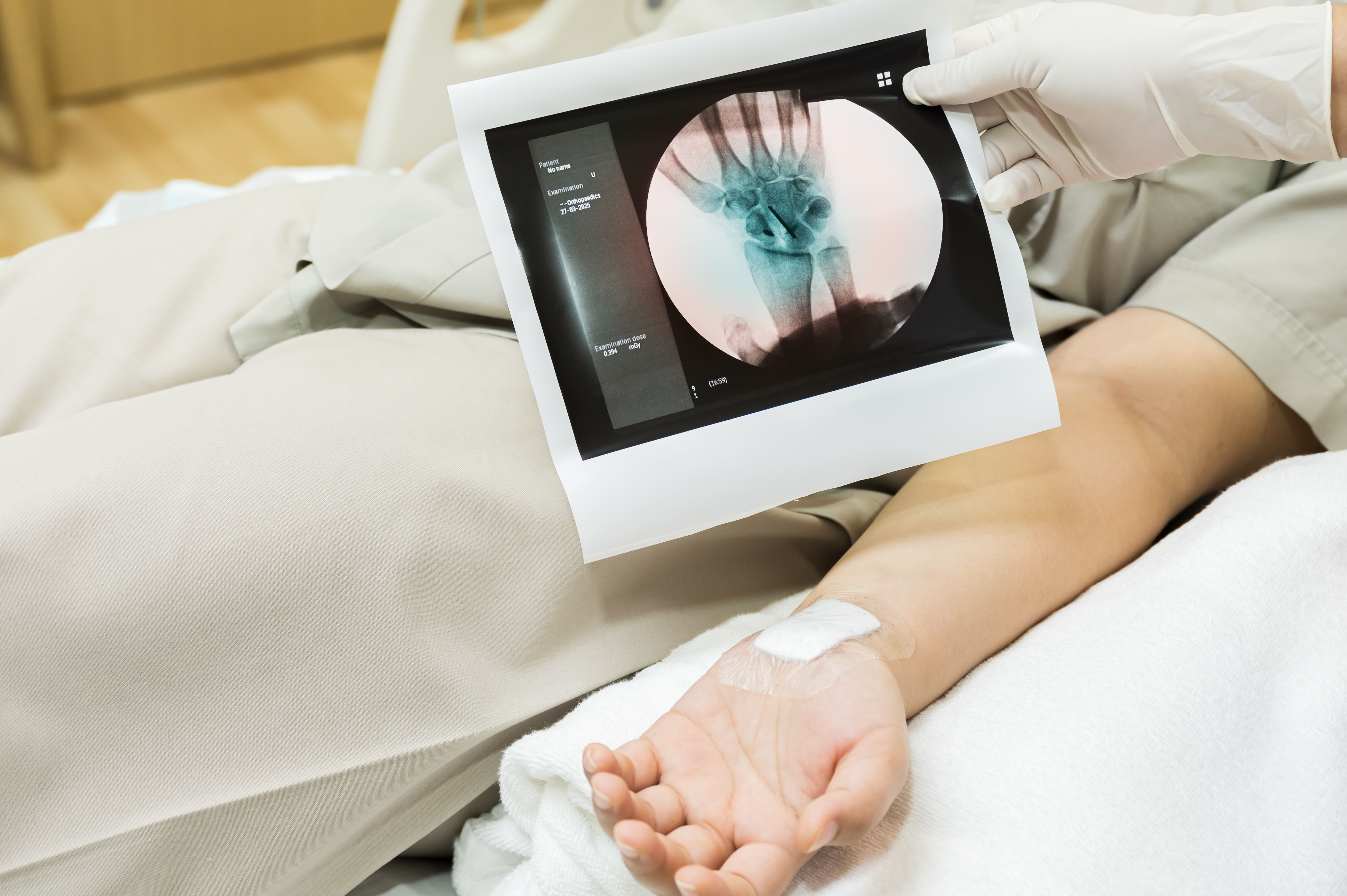
Wrist Arthroscopy
ACL Tear Treatment
Rotator Cuff Tear
Meniscus Transplant
Media
Picture Gallery
Testimonials
Contact
📅 Book Appointment