Dubai, UAE +971 56 774 1007 WhatsApp
Home About
All Services ACL Surgery PCL Surgery Knee Arthroscopy Shoulder Arthroscopy Ankle Arthroscopy Elbow Arthroscopy Hip Arthroscopy Wrist Arthroscopy ACL Tear Treatment Rotator Cuff Tear Meniscus Transplant
Media
Picture Gallery Testimonials Contact 📅 Book Appointment
RC
Shoulder Surgery · Dubai

Rotator Cuff Tear Treatment

Expert diagnosis and arthroscopic repair of rotator cuff tears in Dubai — performed by British-trained FRCS orthopaedic and sports surgeon Dr. Mohammad Ashfaq Konchwalla at Medcare Hospital, Dubai.

Arthroscopic Repair Partial & Full Thickness Tears Supraspinatus Tear Double-Row Fixation Acromioplasty Same-Day Discharge
25+
Years Experience
60–90
Min Procedure
48h
Appointment Wait
FRCS
Royal College Certified
Understanding Rotator Cuff Tears

Shoulder Tendon Injuries Treated with Precision & Care

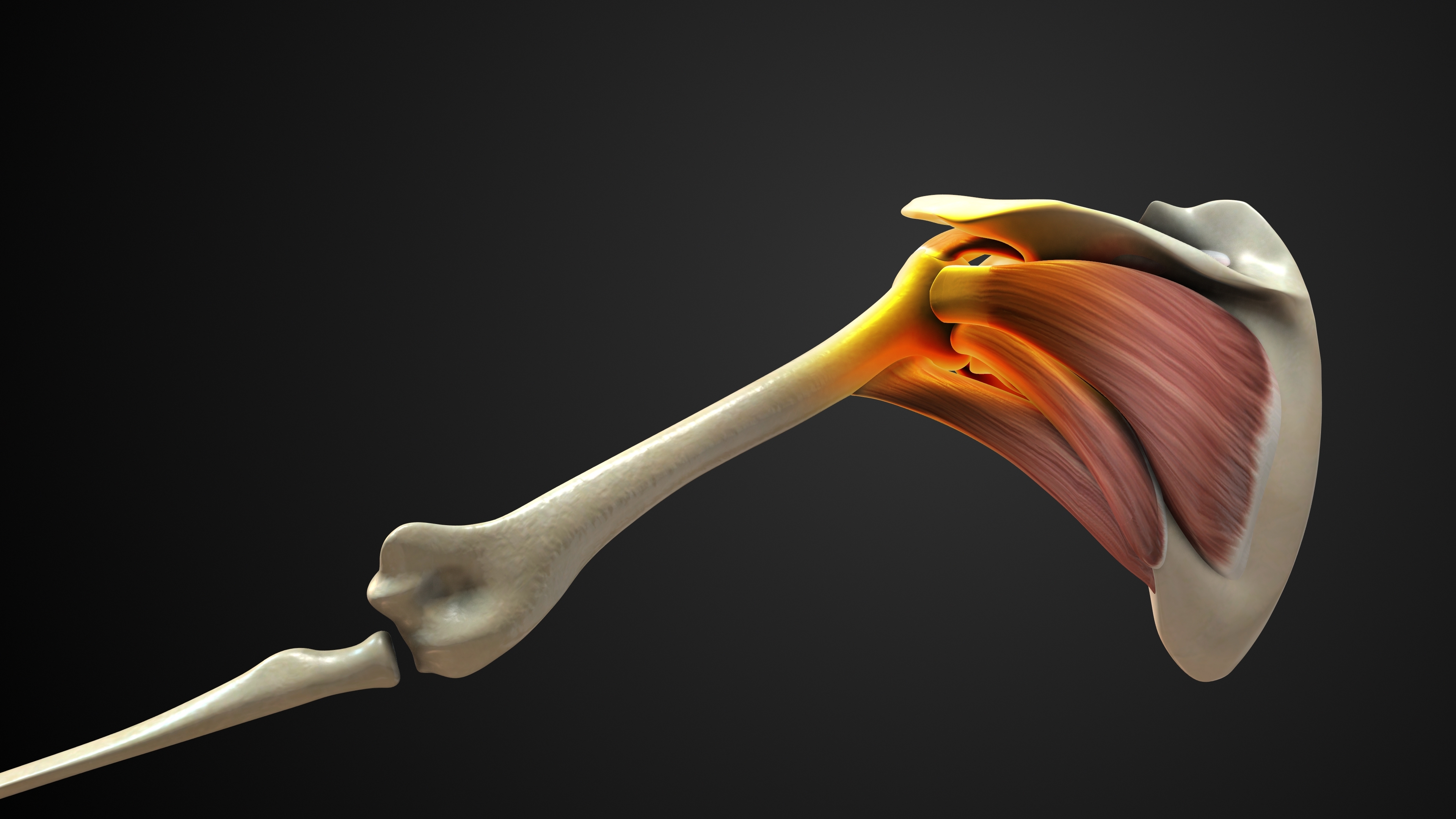
The rotator cuff is a group of four muscles and their associated tendons — the supraspinatus, infraspinatus, teres minor, and subscapularis — that collectively wrap around the head of the humerus, holding it firmly within the shallow glenoid socket of the shoulder blade. The rotator cuff is the essential dynamic stabiliser of the shoulder joint: it centres the humeral head during every movement of the arm, enables the full range of shoulder motion, and generates the force required for lifting, throwing, pushing, and pulling. Without a functioning rotator cuff, the shoulder cannot work normally.

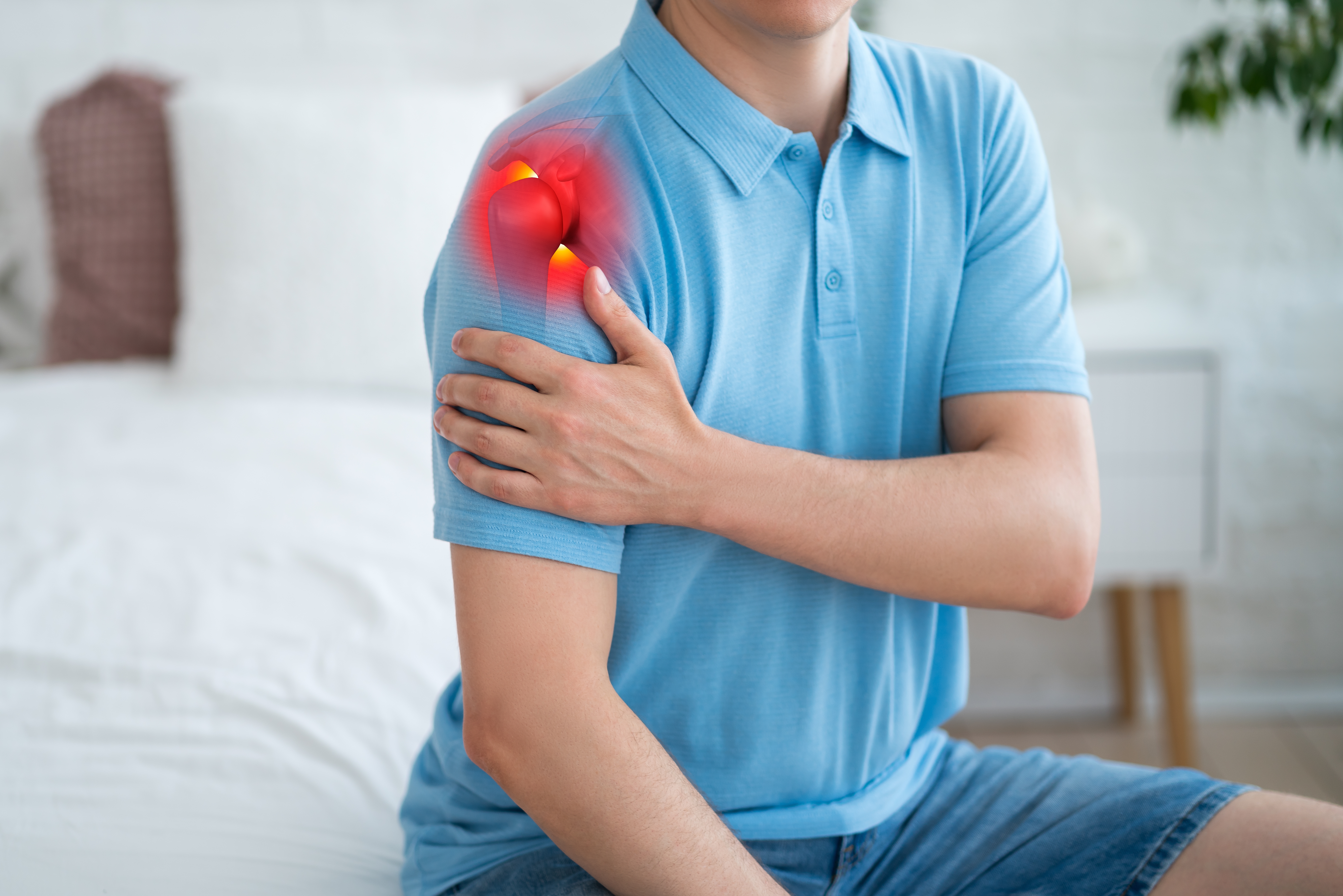
A rotator cuff tear occurs when one or more of these tendons is partially or completely disrupted — either through a sudden traumatic event or through gradual degenerative wear over time. Rotator cuff tears are among the most common causes of shoulder pain and disability in adults, affecting athletes across all sports, active individuals, and older patients whose tendons have weakened with age. The supraspinatus tendon is the most commonly torn — accounting for the vast majority of rotator cuff injuries — due to its anatomical position under the acromion, where it is exposed to impingement with every overhead movement.

Dr. Konchwalla's extensive shoulder surgery expertise, FRCS qualifications from three Royal Colleges, and over two decades of specialist practice in Dubai make him the first choice for patients seeking definitive rotator cuff tear treatment. Whether your injury is a minor partial thickness tear amenable to physiotherapy, or a large full-thickness complete tear requiring arthroscopic repair with double-row suture anchor fixation, Dr. Konchwalla will design a precise, evidence-based treatment plan tailored to your specific tear, your age, your sport, and your recovery goals.

Rotator Cuff Tear Treatment Dubai — Dr. Konchwalla
Procedure Type
Arthroscopic Rotator Cuff Repair
Minimally invasive keyhole surgery through 3–4 small portals. Suture anchors reattach the torn tendon to the greater tuberosity using single-row or double-row fixation.
Duration
60 – 90 Minutes
Simple partial thickness repairs take 45–60 minutes; large full-thickness tears with acromioplasty and double-row repair require 75–90 minutes.
Anaesthesia
General or Regional Anaesthesia
Interscalene nerve block provides excellent intra- and post-operative pain control and reduces the requirement for general anaesthetic agents, enabling a comfortable same-day recovery.
Hospital Stay
Day Case Surgery
Arthroscopic rotator cuff repair at Medcare Hospital Dubai is routinely performed as same-day surgery. Most patients are discharged within a few hours with an arm sling and aftercare instructions.
Post-Op Support
Arm Sling 4–6 Weeks
The arm is protected in a body-position sling to allow tendon-to-bone healing. Structured physiotherapy begins at 6 weeks, with return to sport targeted at 4–6 months.
Consultant Surgeon
Dr. Mohammad Ashfaq Konchwalla
FRCS (Eng), FRCS (Glas), FRCS (Tr & Ortho)
Location
Medcare Hospital, Dubai
22A Street, From Sheikh Zayed Road, 2nd Interchange, Dubai.
Book Consultation
Appointments within 48 hours
WhatsApp or call to schedule your consultation.
Shoulder Anatomy

The Four Muscles of the Rotator Cuff

The rotator cuff comprises four distinct muscles, each with a specific mechanical role that is critical to normal shoulder function. All four originate from the scapula and insert via their tendons onto the greater or lesser tuberosity of the humerus. Together they form a musculotendinous envelope around the glenohumeral joint — providing dynamic stability, compressing the humeral head into the glenoid, and enabling the extraordinary range of motion that makes the shoulder the most mobile joint in the body.

Initiates shoulder abduction (lifting the arm away from the body) and is the most commonly injured tendon. Lies in the supraspinous fossa of the scapula and inserts on the superior greater tuberosity.
The primary external rotator of the shoulder, essential for throwing, serving, and overhead sports. Originates in the infraspinous fossa and inserts on the middle greater tuberosity.
Assists the infraspinatus in external rotation and depresses the humeral head during abduction. Originates from the lateral scapular border and inserts on the inferior greater tuberosity.
The largest and strongest rotator cuff muscle; the principal internal rotator of the shoulder and a primary anterior stabiliser. Inserts via a broad tendon onto the lesser tuberosity of the humerus.
Why Arthroscopic Repair?

Advantages of Minimally Invasive Rotator Cuff Surgery

Arthroscopic rotator cuff repair by Dr. Konchwalla offers significant clinical advantages over traditional open shoulder surgery — less tissue disruption, faster healing, and a more complete diagnostic assessment of the entire shoulder joint at the time of repair.

Arthroscopy provides a full 15-point inspection of the entire shoulder joint — detecting associated labral tears, biceps pathology, cartilage damage, and SLAP lesions that an open approach cannot visualise, ensuring no concurrent pathology is missed.
Small portal incisions — 5–8 mm — preserve the deltoid muscle, the main covering muscle of the shoulder that would be detached and repaired in traditional open surgery. Preserving the deltoid accelerates recovery and reduces post-operative pain substantially.
Arthroscopic rotator cuff repair is performed as day-case surgery at Medcare Hospital Dubai. No overnight admission is required. Patients return home the same day with their arm in a sling, a prescription for pain relief, and a personalised aftercare plan.
Reduced operative trauma means physiotherapy can commence earlier, post-operative pain resolves faster, and patients regain shoulder movement more quickly compared to open surgery. Return to work, sport, and daily activities is accelerated.
Dr. Konchwalla employs double-row suture anchor fixation for large and massive tears — creating a broader tendon-to-bone contact area with superior biomechanical strength compared to single-row repair. This reduces re-tear risk and improves the long-term durability of the repair.
Types of Rotator Cuff Tears

Understanding Your Rotator Cuff Injury

Rotator cuff tears vary widely in their location, depth, size, and underlying cause. Accurate classification is essential to determine the correct treatment — from conservative physiotherapy for minor partial tears to surgical repair for complete full-thickness injuries.

Supraspinatus Tear
The supraspinatus is the most frequently torn rotator cuff tendon, accounting for over 90% of all rotator cuff injuries. It runs horizontally across the top of the shoulder beneath the acromion, making it vulnerable to repetitive impingement and degenerative wear. Supraspinatus tears cause painful arc syndrome — pain in the 60–120° range of shoulder elevation — weakness lifting the arm out to the side, and night pain that disturbs sleep. Small partial supraspinatus tears in older patients with good function can be managed with physiotherapy and subacromial injection. Larger partial tears (greater than 50% tendon thickness) and full-thickness tears in active patients require arthroscopic repair using suture anchors — either single-row or the stronger double-row technique that provides a larger footprint repair with superior biomechanical fixation.
At a Glance
Prevalence
Over 90% of rotator cuff injuries
Key Symptom
Painful arc 60–120°
Repair Technique
Single-row or double-row suture anchors
Infraspinatus & Teres Minor Tears
The infraspinatus and teres minor are the primary external rotators of the shoulder — essential for throwing actions, racket sports, and overhead movements. Isolated infraspinatus tears most commonly occur in overhead athletes and throwing sports specialists, often resulting from posterior shoulder impingement or repetitive eccentric loading of the posterior cuff during deceleration. They cause posterior shoulder pain, painful external rotation, and a dramatic reduction in throwing velocity. Teres minor injuries are rarer but frequently occur in conjunction with infraspinatus tears in massive posterior rotator cuff injuries. Both tendons are repaired arthroscopically using precision suture anchor techniques, restoring the posterior cuff function critical for overhead athletic performance.
At a Glance
Function
Primary external rotators
Key Symptom
Posterior pain, reduced throwing velocity
Common In
Overhead athletes, throwing sports
Subscapularis Tear
The subscapularis is the largest and most powerful rotator cuff muscle — the principal internal rotator of the shoulder — and tears to this tendon are frequently missed or misdiagnosed. Subscapularis tears occur most commonly following anterior shoulder dislocation, a sudden forceful external rotation with the arm abducted, or a direct impact to the front of the shoulder. They cause anterior shoulder pain, weakness with internal rotation and pressing movements, a positive bear-hug test, and a feeling of shoulder instability. Isolated subscapularis tears are repaired arthroscopically through the anterior portals, with meticulous identification and repair of both the superior and inferior components of the tendon. Associated biceps tendon pathology — very commonly present alongside subscapularis tears — is simultaneously addressed.
At a Glance
Function
Principal internal rotator
Key Symptom
Anterior pain, positive bear-hug test
Common Cause
Anterior dislocation, forced external rotation
Massive Rotator Cuff Tears
A massive rotator cuff tear is defined as a complete tear involving two or more tendons, or a single tear with a transverse diameter exceeding 5 cm. Massive tears most commonly affect the supraspinatus and infraspinatus together — resulting in severe loss of shoulder strength, an inability to actively elevate the arm, and progressive glenohumeral arthritis if untreated. The treatment of massive tears is complex: tendon retraction, muscle atrophy, and fatty infiltration must all be assessed on MRI before surgery. Arthroscopic repair with margin convergence stitches and double-row fixation is the preferred approach where tissue quality permits. In irreparable massive tears, superior capsule reconstruction or tendon transfer procedures — including latissimus dorsi transfer — restore shoulder function. Dr. Konchwalla's extensive experience with complex shoulder reconstruction makes him ideally placed to manage these challenging injuries.
At a Glance
Definition
2+ tendons or >5 cm diameter
Key Symptom
Severe weakness, inability to elevate arm
Repair Technique
Margin convergence + double-row fixation
Partial Thickness Rotator Cuff Tears
Partial thickness tears involve damage to a proportion of the rotator cuff tendon fibres without a complete full-thickness defect through the tendon. They are classified by location — articular surface partial tears (PASTA lesions), bursal surface partial tears, or intratendinous partial tears — and by the percentage of tendon thickness involved. Articular-surface partial tears affecting more than 50% of tendon thickness (high-grade PASTA lesions) have poor healing potential and are best treated surgically. Lower-grade partial tears (less than 50%) in young active patients are treated with arthroscopic débridement of frayed tissue and subacromial decompression (acromioplasty) where there is impingement. Careful classification of partial tear grade and location is essential to avoid under- or over-treatment.
At a Glance
Types
Articular (PASTA), bursal, intratendinous
Surgery Threshold
>50% tendon thickness
Conservative Option
Débridement + acromioplasty
Rotator Cuff Tears with Biceps Pathology
The long head of the biceps tendon (LHBT) runs through the bicipital groove of the humerus and has its attachment at the superior labrum and supraglenoid tubercle, making it intimately related to the rotator cuff — particularly the subscapularis and supraspinatus. Biceps tendon pathology — including biceps tendinopathy, partial tears, SLAP tears, and biceps instability — is found concurrently in up to 40% of rotator cuff repairs. Untreated biceps pathology is a significant cause of persistent anterior shoulder pain following rotator cuff repair. Dr. Konchwalla performs thorough assessment of the biceps tendon at every shoulder arthroscopy, treating biceps pathology concurrently with rotator cuff repair through arthroscopic biceps tenodesis (reattachment to the humerus) or tenotomy as clinically indicated.
At a Glance
Prevalence
Up to 40% of rotator cuff repairs
Key Symptom
Persistent anterior shoulder pain
Treatment
Biceps tenodesis or tenotomy
Surgical Process

Your Rotator Cuff Repair — Step by Step

Dr. Konchwalla follows a systematic, evidence-based surgical protocol for every rotator cuff repair — ensuring complete assessment of the shoulder joint, precise tendon reattachment, and comprehensive treatment of any associated pathology.

01

Assessment & Portal Placement

Start
Under general or interscalene regional anaesthesia, the patient is positioned in the beach-chair position. Three to four arthroscopic portals of 5–8 mm are carefully placed around the shoulder. Fluid distends the glenohumeral joint. The arthroscope is introduced and a systematic 15-point inspection of the entire shoulder joint is performed — assessing the rotator cuff from the articular side, the biceps tendon and labrum, the glenohumeral ligaments, the cartilage surfaces, and the axillary pouch.
Beach-Chair Position 3-4 Portals 15-Point Inspection
02

Subacromial Assessment & Acromioplasty

Preparation
The arthroscope is moved into the subacromial space, and the bursal surface of the rotator cuff is inspected. Any thickened, inflamed subacromial bursa is removed using a motorised shaver (bursectomy) to provide optimal visualisation of the cuff from above. Where there is subacromial impingement due to a hooked (Type 3) acromion or acromioclavicular joint spurs, an arthroscopic acromioplasty is performed using a small bone shaver to create a flat acromion — reducing impingement and providing a better healing environment for the repaired tendon.
Bursectomy Acromioplasty Impingement Clearance
03

Tendon Mobilisation & Footprint Preparation

Preparation
The torn tendon edges are mobilised by carefully releasing any adhesions that have developed since the tear, allowing the tendon to be brought back to its anatomical insertion on the greater tuberosity. The footprint of the greater tuberosity — the bone surface to which the tendon will be reattached — is prepared by removing any remaining scar tissue, decorticated, and lightly abraded to create a vascular, bleeding bone bed that promotes biological healing and tendon integration.
Adhesion Release Footprint Decortication Bleeding Bone Bed
04

Suture Anchor Repair & Closure

Core Procedure
High-strength suture anchors (titanium or bioabsorbable) are inserted into the prepared greater tuberosity footprint. The sutures from these anchors are passed through the tendon using precision shuttling instruments and tied arthroscopically to pull the tendon down firmly onto the bone. For larger tears, double-row fixation with medial and lateral rows of anchors is used — maximising the contact area between tendon and bone for superior biological healing. Associated biceps pathology is treated concurrently. The portals are closed with a single absorbable suture and an arm sling is applied before waking.
Suture Anchors Double-Row Fixation Biceps Treatment Fine Sutures
60–90 Min
Procedure
3-4
Portals
Same Day
Discharge
4-6 mo
Return to Sport
Book Consultation
Recovery Timeline

What to Expect After Rotator Cuff Repair

Recovery after rotator cuff repair surgery is a structured, progressive process. The tendon requires time to heal biologically to the bone — and respecting this healing timeline is critical to achieving a durable, functional outcome. Dr. Konchwalla's team of specialist shoulder physiotherapists supervise your rehabilitation at every stage.

90%+
Return-to-Sport Rate
Week 1–2
Phase 1
Rest & Swelling Control
The arm is kept in an abduction or body-position sling at all times. Ice packs and prescribed anti-inflammatory pain relief manage post-operative swelling and discomfort. The wound is reviewed at 10–14 days. Pendulum exercises begin from Day 1 to prevent shoulder stiffness.
Week 2–6
Phase 2
Sling Protection & Passive Physiotherapy
The arm sling is maintained to protect the healing tendon-to-bone repair. Supervised physiotherapy focuses on passive and assisted range-of-motion exercises — gentle shoulder elevation, external rotation, and cross-body stretching — to restore flexibility without loading the repair. Elbow, wrist, and hand mobility exercises prevent stiffness in the distal arm.
Week 6–12
Phase 3
Active Range of Motion
The sling is discontinued at 6 weeks once tendon-to-bone healing is confirmed. Active-assisted and then fully active shoulder range-of-motion exercises commence. Physiotherapy sessions are increased in frequency and intensity. Rotator cuff muscle activation and scapular stabilisation exercises begin. Driving is permitted from 6–8 weeks.
Month 3–4
Phase 4
Strengthening Programme
Progressive rotator cuff and periscapular strengthening exercises are introduced using resistance bands, weights, and gym-based protocols. Functional shoulder exercises replicate sport-specific movement patterns. Upper body resistance training — excluding overhead pressing — is resumed. Shoulder strength and proprioception are systematically rebuilt.
Month 4–6
Phase 5
Return to Sport
Sport-specific loading, throwing progressions, and overhead training are introduced under physiotherapy guidance. Return to non-contact recreational sport is typically achieved at 4–5 months. Return to full competitive sport — including cricket, tennis, swimming, CrossFit, and rugby — at 5–6 months, subject to objective strength testing confirming at least 90% symmetry with the contralateral shoulder.
Your Surgeon

Expert Rotator Cuff Care in Dubai

Why Dr. Konchwalla?
Qualifications
FRCS (Eng), FRCS (Glas), FRCS (Tr & Ortho) — Fellow of three Royal Colleges, trained at King's College London.
Experience
25+ years of specialist shoulder surgery practice in Dubai — one of the region's most experienced shoulder surgeons.
Approach
Conservative-first philosophy — surgery is recommended only when non-surgical management has failed or when the tear demands repair to prevent progression.
Personalised Rehab
Sport-specific rehabilitation protocols tailored to your sport, your position, and your competitive level — ensuring a safe, complete return to performance.
"A rotator cuff tear left untreated does not simply become painful — it becomes larger, and the tendon retracts further. Timely repair, when tissue quality is optimal and muscle atrophy is minimal, delivers the best possible outcome. Every week of delay matters for the athlete who wants to return to full performance."
Dr. Mohammad Ashfaq Konchwalla — FRCS (Eng), FRCS (Glas), FRCS (Tr & Ortho)

Rotator Cuff Tears in Athletes

Rotator cuff injuries are among the most common sport-limiting shoulder injuries in Dubai's active population. Dr. Konchwalla regularly treats elite and recreational athletes across a wide range of sports with sport-specific repair and return-to-play protocols.

🏏
Cricket
Supraspinatus and infraspinatus tears from high-volume bowling and throwing — particularly fast bowlers with repetitive overhead delivery action. Posterior cuff injuries from deceleration forces and dive catches. Subscapularis injuries from sudden bat impact.
🎾
Tennis & Racket Sports
Supraspinatus impingement and partial tears from the overhead serve and smash. Infraspinatus tears from the deceleration phase of the service action. Posterior cuff overuse from high-volume competitive training leading to progressive rotator cuff degeneration.
🏊
Swimming
Swimmer's shoulder — supraspinatus and infraspinatus impingement and partial tearing from the repetitive overhead pulling action of freestyle, butterfly, and backstroke. Internal impingement at the posterior-superior cuff from the extreme shoulder positions of competitive swimming.
🏋️
CrossFit & Weightlifting
Supraspinatus tears from overhead pressing, kipping pull-ups, and barbell snatches. Subscapularis injuries from bench press and ring dip loading. Rotator cuff impingement from high-volume overhead programming in CrossFit athletes without adequate shoulder stability and scapular control.
FAQs

Frequently Asked Questions

Common questions about rotator cuff tear treatment and repair surgery in Dubai — answered by Dr. Konchwalla's team at Dubai Sports Surgery.

Partial rotator cuff tears in older, lower-demand patients can improve significantly with physiotherapy and subacromial injection. However, rotator cuff tendons have poor intrinsic healing capacity due to limited vascularity at the tendon-bone junction. Complete full-thickness tears do not heal spontaneously and will typically enlarge over time. Dr. Konchwalla assesses each patient individually, and surgical repair is recommended for active patients, athletes, and patients with large or full-thickness tears where conservative treatment has failed.
Recovery follows a structured timeline. Arm sling: 4–6 weeks. Passive physiotherapy: weeks 2–6. Active range of motion: from 6 weeks. Strengthening programme: months 3–4. Return to recreational sport: 4–5 months. Return to competitive and overhead sport: 5–6 months. Large and massive tears require a longer protocol. Tendon-to-bone healing is a biological process that cannot be rushed — respecting the timeline is essential to prevent re-tear.
Double-row repair uses two rows of suture anchors — a medial row and a lateral row — to reattach the torn rotator cuff tendon across its full anatomical footprint on the greater tuberosity. Compared to single-row repair, double-row fixation creates a larger contact area between tendon and bone, provides higher initial fixation strength, and has been shown to result in better biological healing. Dr. Konchwalla uses double-row techniques for large and massive tears where the footprint reconstruction is critical to long-term durability.
Diagnosis combines clinical examination, imaging, and in some cases, diagnostic arthroscopy. Dr. Konchwalla performs specific clinical tests — Jobe's test, the drop arm sign, Hawkins-Kennedy, and the Neer impingement sign. MRI is the gold standard investigation, providing detailed assessment of tear size, tendon retraction, muscle quality, and fatty infiltration. Musculoskeletal ultrasound is a useful dynamic adjunct. Diagnostic shoulder arthroscopy allows direct visualisation of the cuff when imaging is inconclusive.
Yes. Arthroscopic rotator cuff repair at Medcare Hospital Dubai is routinely performed as same-day day-case surgery under general or interscalene regional anaesthesia. Most patients are discharged within 3–4 hours of the procedure with an arm sling, pain relief, and written aftercare instructions. Overnight hospital admission is rarely required and is reserved for patients with significant medical comorbidities or those undergoing complex massive cuff reconstruction.
Untreated full-thickness rotator cuff tears progressively enlarge over time — retract further from the bone, develop muscle atrophy, and undergo fatty infiltration of the rotator cuff muscle bellies. This fatty infiltration is irreversible and negatively affects both the technical quality of surgical repair and the functional outcome. Massive, chronically retracted tears may become irreparable. Prolonged tears also result in superior migration of the humeral head, cartilage damage, and ultimately rotator cuff arthropathy. Early repair, when tissue quality is optimal, delivers the best outcomes.
Driving is not permitted while the arm sling is being worn — typically for the first 6 weeks following surgery. After the sling is removed and physiotherapy has restored adequate shoulder range of motion, strength, and reaction time, driving can be resumed — usually at 6–8 weeks for automatic vehicles and 8–10 weeks for manual vehicles. Dr. Konchwalla will confirm clearance to drive at your 6-week post-operative review.
Rotator cuff tears occur through two mechanisms. Acute traumatic tears result from a fall on an outstretched arm, a direct blow to the shoulder, a sudden forced external rotation, or a rapid overhead jerk — such as catching a heavy falling object. Degenerative tears develop gradually from age-related tendon degeneration, repetitive overhead loading, and cumulative impingement wear — most commonly affecting the supraspinatus tendon in patients over 40. Risk factors include advancing age, overhead sport participation, previous shoulder impingement, diabetes, corticosteroid use, and smoking, which impairs tendon vascularity and healing capacity.
Book the Best Dubai Sports Surgeon

Consult Dr. Ashfaq Konchwalla
Reclaim Your Peak

Book a consultation with Dubai’s top sports surgery expert — British-trained orthopaedic & sports surgery specialist. Take the first step toward a full, confident recovery. Appointments available within 48 hours.

Location
Dubai, United Arab Emirates
Clinic Hours
Sunday – Thursday  9:00 – 17:00
Emergencies
Available on Request